Chronic & Autoimmune Disorders
Psoriasis
At MD Claiborne Dermatology, we specialize in personalized psoriasis care and are one of the few practices offering full-body narrowband UVB (nbUVB) phototherapy, a safe, non-immunosuppressive treatment for plaque psoriasis.
For moderate-to-severe psoriasis, our dermatologists utilize advanced biologic therapies that calm overactive immune responses and clear skin effectively. Common biologic classes include:
Anti-TNFs (Humira®, Enbrel®, Remicade®, Cimzia®)
Anti-IL-17s (Cosentyx®, Taltz®, Siliq®)
Anti-IL-23s (Skyrizi®, Tremfya®)
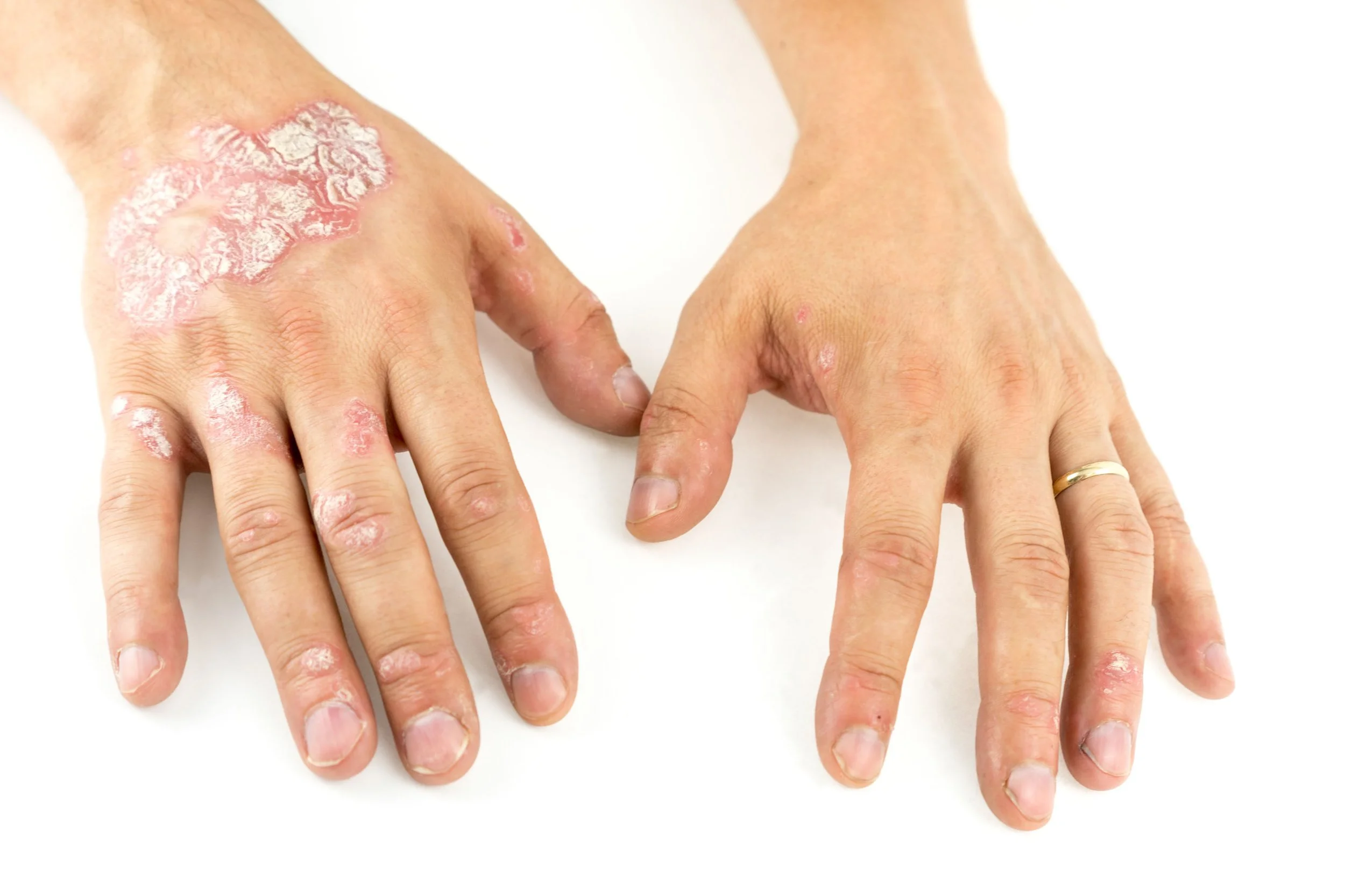
Psoriasis is a chronic autoimmune skin condition that speeds up skin-cell growth, causing red, thickened, scaly patches most often on the elbows, knees, scalp, and lower back. The condition is non-contagious but can vary from mild flare-ups to severe, full-body involvement.
While there’s no cure, psoriasis often improves with sunlight, warm weather, or targeted therapy—and some patients experience years of remission. Triggers may include cold, dry climates, stress, or illness. Psoriasis affects more than 8 million Americans of all ages and backgrounds, typically appearing in young adulthood.
Dr. Cole Claiborne lead ongoing clinical research trials in partnership with the University of Pennsylvania and the National Psoriasis Foundation, bringing cutting-edge psoriasis treatments to patients across Louisiana.

Major Types of Psoriasis
Plaque Psoriasis: The most common form, appearing as raised, red, inflamed patches covered with silvery-white scales. It commonly affects the elbows, knees, and scalp but can occur anywhere on the body
Guttate Psoriasis: Characterized by small, red, scaly spots that appear suddenly. It is more common in children and young adults and can be triggered by a bacterial infection, like strep throat.
Inverse Psoriasis: Occurs in skin folds, such as the armpits, groin, and under the breasts. It appears as smooth, shiny, red or darker patches of skin rather than with scales.
Pustular Psoriasis: A less common type that presents with pus-filled blisters (pustules) surrounded by red skin. The pustules are not infectious, and this type can be localized to the hands and feet or more widespread.
Erythrodermic Psoriasis: A severe and rare form that can cover large areas of the body. It causes intense redness and peeling of the skin and is considered a medical emergency because it can lead to severe illness.

Eczema (Dermatitis)
Eczema, also known as dermatitis, is a common inflammatory skin condition that causes redness, itching, swelling, and flaking. In acute stages, eczema may blister or ooze; in chronic cases, skin becomes thickened, leathery, and darker in tone.
At MD Claiborne Dermatology, our offices offer phototherapy, a safe, non-immunosuppressive treatment for eczema in patients of all ages. Severe or persistent cases may require systemic medications such as methotrexate or cyclosporine.
Breakthrough biologic therapies, including Dupixent® (dupilumab) and oral JAK Inhibitors like RINVOQ, and Cibinqo have revolutionized care for patients with severe atopic dermatitis. Dr. Cole Claiborne participated in the clinical trials for Dupixent and provides expert assessment to determine whether it’s right for you.
Common Types of Eczema
Atopic Dermatitis: The most common and chronic form of eczema. It can cause dry, itchy, and inflamed patches of skin.
Contact Dermatitis: Occurs when skin reacts to an irritant (like soap or detergent) or an allergen (like nickel or poison ivy).
Dyshidrotic Eczema: Characterized by small, itchy blisters on the hands, fingers, and feet. It's often triggered by stress or contact with certain metals.
Nummular Eczema: Presents as distinct, coin-shaped or oval patches that are often itchy and can sometimes ooze.
Seborrheic Dermatitis: A chronic type that affects oily areas such as the scalp, face, and upper chest.
Statis Dermatitis: Occurs in people with poor circulation, most often in the lower legs. It results in swelling, redness, and dry, itchy skin.
Neurodermatitis: This type involves a localized, intensely itchy patch of skin that can become leathery from scratching.

Vitiligo
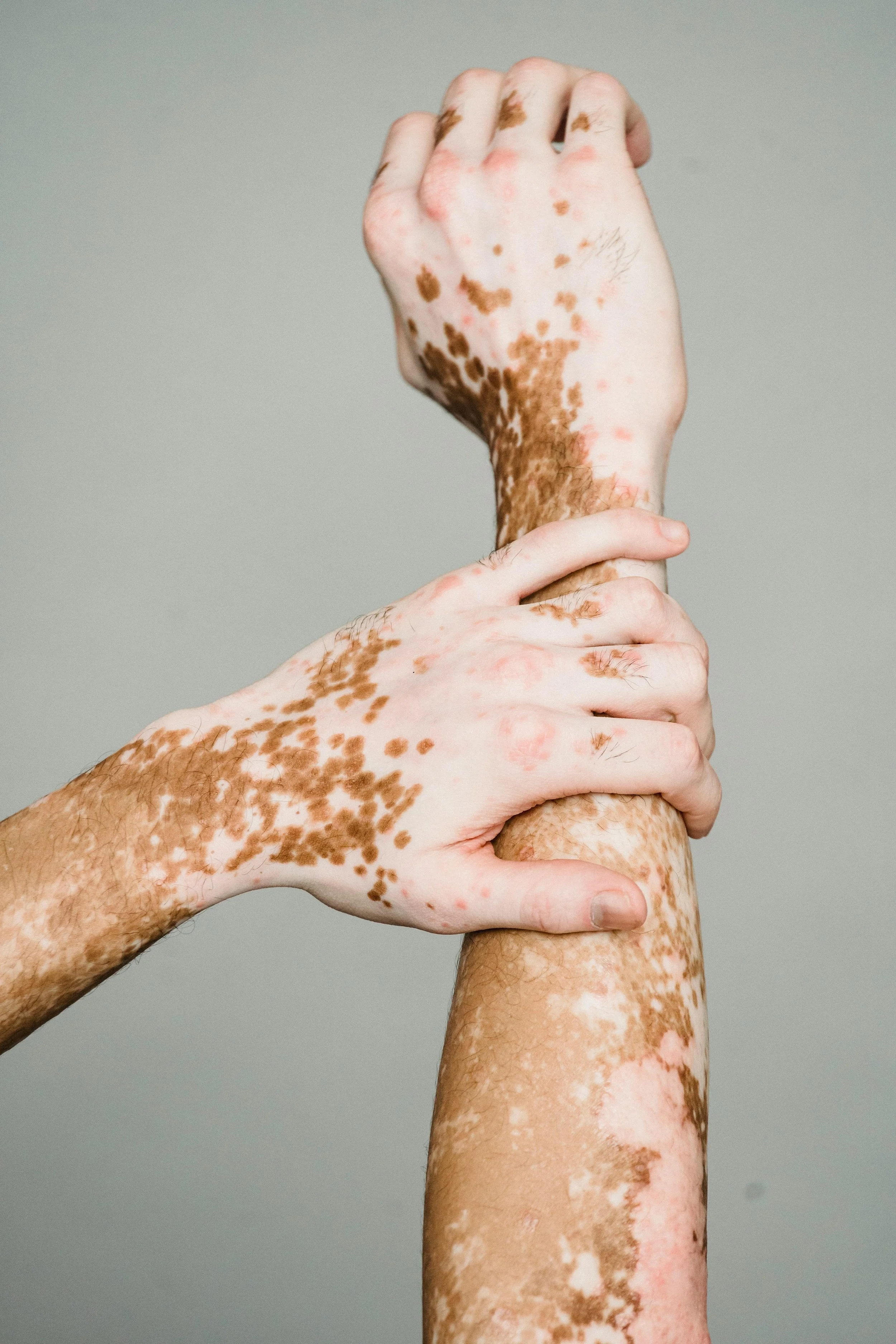
Vitiligo is an autoimmune skin condition that causes loss of pigmentation, resulting in light or white patches that can appear anywhere on the body. The condition occurs when melanocytes—the pigment-producing cells in the skin—are destroyed. The exact cause remains unclear, but factors like autoimmune activity, stress, or genetic predisposition may contribute.
Vitiligo affects people of all races and most commonly begins between ages 20 and 40. In some cases, skin pigment may partially or fully return over time; in others, the color loss can be long-lasting without treatment.
At MD Claiborne Dermatology, our board-certified dermatologists specialize in advanced therapies to restore or even out skin tone, including:
Ultraviolet (UV) light therapy to stimulate pigment cell activity
Topical corticosteroids or calcineurin inhibitors to control immune response
Skin grafting for stable, localized patches
Depigmentation therapy for widespread pigment loss
Early evaluation and customized treatment can significantly improve results and confidence.
Keloids are firm, raised overgrowths of scar tissue that develop after skin injury, burns, piercings, or surgery. They can appear as single nodules or clusters, and may cause itching, pain, or restricted movement depending on their size and location. While keloids are more common among individuals of African-American and Mediterranean descent, they can occur in anyone.
After an injury, the body produces collagen to heal the skin. In some people, this process becomes overactive, creating excess fibrous tissue that extends beyond the original wound.
Because keloids have a high rate of recurrence, treatment must be carefully planned and managed by a board-certified dermatologist. Options include:
Intralesional steroid injections to flatten and soften the scar
Surgical excision combined with post-procedure therapy to prevent regrowth
Cryotherapy, laser therapy, or radiation treatments in resistant cases
Chemotherapeutic agents such as 5-fluorouracil (5-FU) for severe or recurrent keloids
Keloids & Scars
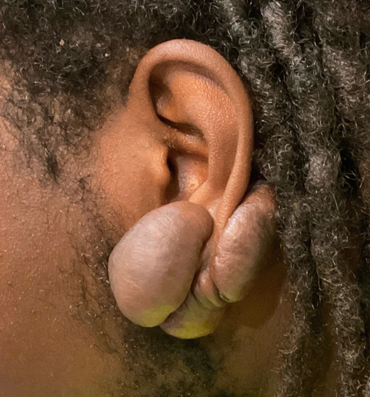
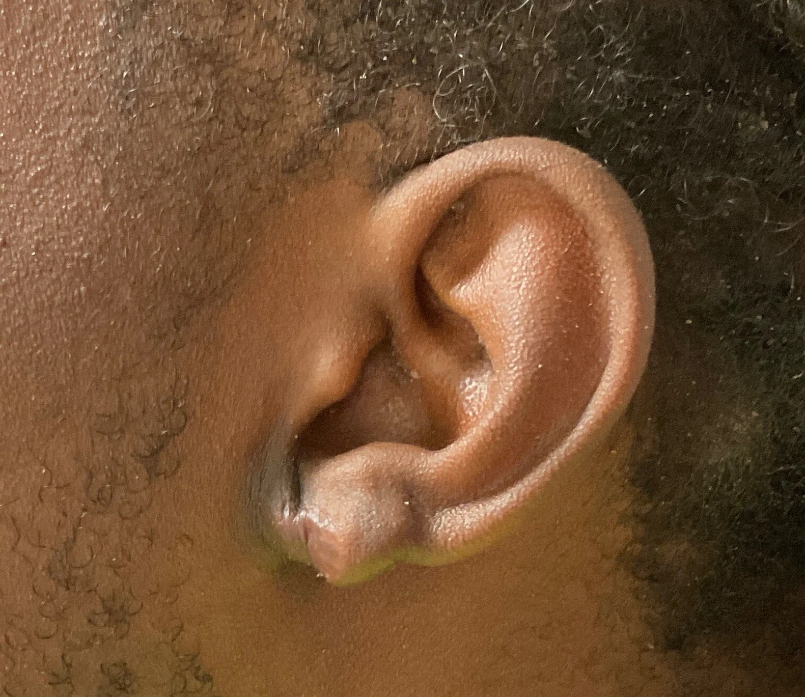
Keloid Before & After

At MD Claiborne Dermatology, Dr. Claiborne uses the T.R.U.E. Test™ (Thin-layer Rapid Use Epicutaneous Test) to identify up to 35 common contact allergens.
This topical “patch test” safely detects skin allergies without breaking the skin—unlike the “prick” or “scratch” tests used for respiratory or food allergies. Note: this test is not used to detect allergens associated with anaphylaxis.
Once the allergen is identified, treatment may include topical or oral corticosteroids, emollients, and avoidance strategies to prevent future reactions.